What is the meaning of the term germinate? to store food to start growth to make eggs to make sperm
In 1 respect at least, eggs are the about remarkable of animate being cells: one time activated, they tin give rise to a complete new private within a matter of days or weeks. No other cell in a higher creature has this capacity. Activation is normally the issue of fertilization—fusion of a sperm with the egg. In some organisms, even so, the sperm itself is not strictly required, and an egg can exist activated artificially by a variety of nonspecific chemical or physical treatments. Indeed, some organisms, including a few vertebrates such equally some lizards, normally reproduce from eggs that become activated in the absence of sperm—that is, parthenogenetically.
Although an egg can give rise to every cell type in the adult organism, it is itself a highly specialized jail cell, uniquely equipped for the single function of generating a new private. The cytoplasm of an egg can fifty-fifty reprogram a somatic cell nucleus so that the nucleus can directly the development of a new individual. That is how the famous sheep Dolly was produced. The nucleus of an unfertilized sheep egg was destroyed and replaced with the nucleus of an adult somatic cell. An electrical shock was used to actuate the egg, and the resulting embryo was implanted in the uterus of a surrogate mother. The resulting normal developed sheep had the genome of the donor somatic cell and was therefore a clone of the donor sheep.
In this section, we briefly consider some of the specialized features of an egg earlier discussing how information technology develops to the point of being set up for fertilization.
An Egg Is Highly Specialized for Independent Development, with Large Nutrient Reserves and an Elaborate Coat
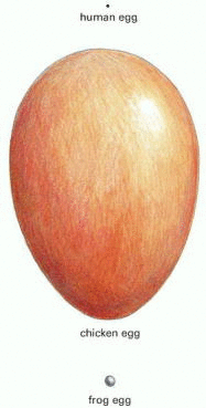
The eggs of most animals are giant single cells, containing stockpiles of all the materials needed for initial development of the embryo through to the stage at which the new individual can brainstorm feeding. Before the feeding phase, the behemothic cell cleaves into many smaller cells, but no net growth occurs. The mammalian embryo is an exception. It can start to grow early by taking up nutrients from the mother via the placenta. Thus, a mammalian egg, although all the same a large jail cell, does not have to exist as large as a frog or bird egg, for example. In general, eggs are typically spherical or ovoid, with a diameter of virtually 0.ane mm in humans and bounding main urchins (whose feeding larvae are tiny), 1 mm to two mm in frogs and fishes, and many centimeters in birds and reptiles (Effigy 20-19). A typical somatic prison cell, by contrast, has a diameter of simply about 10 or 20 μm (Figure 20-20).

Figure 20-19
The bodily sizes of 3 eggs. The human egg is 0.1 mm in diameter.

Figure 20-xx
The relative sizes of various eggs. Sizes are compared with that of a typical somatic prison cell.
The egg cytoplasm contains nutritional reserves in the grade of yolk, which is rich in lipids, proteins, and polysaccharides and is usually contained inside detached structures chosen yolk granules. In some species, each yolk granule is membrane-enclosed, whereas in others information technology is not. In eggs that develop into large animals outside the mother'south body, yolk can business relationship for more than than 95% of the volume of the cell. In mammals, whose embryos are largely nourished past their mothers, there is little, if any, yolk.
The egg glaze is another peculiarity of eggs. It is a specialized form of extracellular matrix consisting largely of glycoprotein molecules, some secreted past the egg and others deposited on it by surrounding cells. In many species, the major coat is a layer immediately surrounding the egg plasma membrane; in nonmammalian eggs, such every bit those of sea urchins or chickens, it is called the vitelline layer, whereas in mammalian eggs it is chosen the zona pellucida (Figure 20-21). This layer protects the egg from mechanical harm, and in many eggs it also acts every bit a species-specific bulwark to sperm, admitting only those of the aforementioned or closely related species.

Figure twenty-21
The zona pellucida. (A) Scanning electron micrograph of a hamster egg, showing the zona pellucida. (B) A scanning electron micrograph of a similar egg in which the zona (to which many sperm are attached) has been peeled back to reveal the underlying plasma (more...)
Many eggs (including those of mammals) comprise specialized secretory vesicles merely under the plasma membrane in the outer region, or cortex, of the egg cytoplasm. When the egg is activated by a sperm, these cortical granules release their contents by exocytosis; the contents of the granules act to alter the egg coat so as to prevent more than 1 sperm from fusing with the egg (discussed beneath).
Cortical granules are normally distributed evenly throughout the egg cortex, just in some organisms other cytoplasmic components accept a strikingly asymmetrical distribution. Some of these localized components later serve to help establish the polarity of the embryo, as discussed in Chapter 21.
Eggs Develop in Stages
A developing egg is called an oocyte. Its differentiation into a mature egg (or ovum) involves a series of changes whose timing is geared to the steps of meiosis in which the germ cells become through their ii last, highly specialized divisions. Oocytes have evolved special mechanisms for arresting progress through meiosis: they remain suspended in prophase I for a prolonged period while the oocyte grows in size, and in many cases they later arrest in metaphase 2 while awaiting fertilization (although they can arrest at diverse other points, depending on the species).
While the details of oocyte development (oogenesis) vary from species to species, the general stages are similar, as outlined in Figure 20-22. Primordial germ cells migrate to the forming gonad to become oogonia, which proliferate by mitosis for a flow before differentiating into primary oocytes. At this stage (usually before nativity in mammals), the first meiotic division begins: the Deoxyribonucleic acid replicates so that each chromosome consists of two sis chromatids, the duplicated homologous chromosomes pair forth their long axes, and crossing-over occurs between nonsister chromatids of these paired chromosomes. After these events, the prison cell remains arrested in prophase of division I of meiosis (in a country equivalent, as we previously pointed out, to a G2 phase of a mitotic sectionalisation wheel) for a menses lasting from a few days to many years, depending on the species. During this long menstruation (or, in some cases, at the onset of sexual maturity), the primary oocytes synthesize a coat and cortical granules. In the case of large nonmammalian oocytes, they also accumulate ribosomes, yolk, glycogen, lipid, and the mRNA that volition later direct the synthesis of proteins required for early embryonic growth and the unfolding of the developmental program. In many oocytes, the intensive biosynthetic activities are reflected in the structure of the chromosomes, which decondense and form lateral loops, taking on a characteristic "lampbrush" appearance, signifying that they are very busily engaged in RNA synthesis (see Figures iv-36 and 4-37).

Figure 20-22
The stages of oogenesis. Oogonia develop from primordial germ cells that migrate into the developing gonad early in embryogenesis. After a number of mitotic divisions, oogonia begin meiotic division I, after which they are called chief oocytes. In mammals, (more than...)
The side by side stage of oocyte development is called oocyte maturation. Information technology usually does non occur until sexual maturity, when the oocyte is stimulated by hormones. Under these hormonal influences, the cell resumes its progress through division I of meiosis. The chromosomes recondense, the nuclear envelope breaks downwardly (this is mostly taken to mark the outset of maturation), and the replicated homologous chromosomes segregate at anaphase I into ii daughter nuclei, each containing half the original number of chromosomes. To end division I, the cytoplasm divides asymmetrically to produce 2 cells that differ profoundly in size: one is a modest polar body, and the other is a big secondary oocyte, the forerunner of the egg. At this stage, each of the chromosomes is still composed of two sister chromatids. These chromatids do non split until partition II of meiosis, when they are partitioned into divide cells, every bit previously described. Later this last chromosome separation at anaphase II, the cytoplasm of the large secondary oocyte once more divides asymmetrically to produce the mature egg (or ovum) and a second small polar torso, each with a haploid set up of single chromosomes (see Effigy twenty-22). Considering of these two asymmetrical divisions of their cytoplasm, oocytes maintain their large size despite undergoing the two meiotic divisions. Both of the polar bodies are small, and they somewhen degenerate.
In about vertebrates, oocyte maturation gain to metaphase of meiosis Two and so arrests until fertilization. At ovulation, the arrested secondary oocyte is released from the ovary and undergoes a rapid maturation stride that transforms information technology into an egg that is prepared for fertilization. If fertilization occurs, the egg is stimulated to complete meiosis.
Oocytes Utilise Special Mechanisms to Grow to Their Large Size
A somatic cell with a diameter of 10–20 μm typically takes most 24 hours to double its mass in preparation for cell division. At this rate of biosynthesis, such a cell would take a very long time to reach the thousand-fold greater mass of a mammalian egg with a diameter of 100 μm. It would accept even longer to attain the million-fold greater mass of an insect egg with a diameter of 1000 μm. Yet some insects live only a few days and manage to produce eggs with diameters even greater than grand μm. It is clear that eggs must accept special mechanisms for achieving their large size.
One simple strategy for rapid growth is to have actress gene copies in the prison cell. Thus, the oocyte delays completion of the first meiotic division so as to grow while it contains the diploid chromosome gear up in duplicate. In this way, it has twice as much Deoxyribonucleic acid available for RNA synthesis as does an average somatic jail cell in the One thousand1 stage of the cell cycle. The oocytes of some species become to fifty-fifty greater lengths to accumulate extra Dna: they produce many extra copies of certain genes. We talk over in Affiliate 6 how the somatic cells of nigh organisms require 100 to 500 copies of the ribosomal RNA genes in lodge to produce plenty ribosomes for protein synthesis. Eggs crave even greater numbers of ribosomes to support protein synthesis during early embryogenesis, and in the oocytes of many animals the ribosomal RNA genes are specifically amplified; some amphibian eggs, for example, contain 1 or 2 meg copies of these genes.
Oocytes may as well depend partly on the synthetic activities of other cells for their growth. Yolk, for example, is ordinarily synthesized outside the ovary and imported into the oocyte. In birds, amphibians, and insects, yolk proteins are fabricated by liver cells (or their equivalents), which secrete these proteins into the claret. Within the ovaries, oocytes take up the yolk proteins from the extracellular fluid by receptor-mediated endocytosis (see Figure 13-41). Nutritive help can also come from neighboring accessory cells in the ovary. These tin can be of two types. In some invertebrates, some of the progeny of the oogonia become nurse cells instead of condign oocytes. These cells usually are continued to the oocyte by cytoplasmic bridges through which macromolecules can pass directly into the oocyte cytoplasm (Figure 20-23). For the insect oocyte, the nurse cells manufacture many of the products—ribosomes, mRNA, protein, and so on—that vertebrate oocytes take to manufacture for themselves.

Figure 20-23
Nurse cells and follicle cells associated with a Drosophila oocyte. The nurse cells and the oocyte arise from a mutual oogonium, which gives rise to one oocyte and 15 nurse cells (only 7 of which are seen in this plane of section). These cells remain (more than...)
The other accessory cells in the ovary that help to nourish developing oocytes are ordinary somatic cells called follicle cells, which are found in both invertebrates and vertebrates. They are arranged every bit an epithelial layer around the oocyte (Figure 20-24, and encounter Figure 20-23), to which they are connected only by gap junctions, which permit the exchange of small molecules but not macromolecules. While these cells are unable to provide the oocyte with preformed macromolecules through these communicating junctions, they may help to supply the smaller precursor molecules from which macromolecules are made. In addition, follicle cells frequently secrete macromolecules that contribute to the egg coat, or are taken up past receptor-mediated endocytosis into the growing oocyte, or human activity on egg cell-surface receptors to control the spatial patterning and axial asymmetries of the egg (discussed in Chapter 21).

Effigy 20-24
Electron micrographs of developing master oocytes in the rabbit ovary. (A) An early stage of primary oocyte development. Neither a zona pellucida nor cortical granules accept developed, and the oocyte is surrounded by a single layer of flattened follicle (more...)
Summary
Eggs develop in stages from primordial germ cells that migrate into the developing gonad early on in development to become oogonia. Afterward mitotic proliferation, oogonia become primary oocytes, which begin meiotic partition I and and so abort at prophase I for days to years, depending on the species. During this prophase-I arrest period, primary oocytes grow, synthesize a glaze, and accumulate ribosomes, mRNAs, and proteins, often enlisting the help of other cells, including surrounding accessory cells. In the process of maturation, main oocytes complete meiotic division I to form a small polar body and a big secondary oocyte, which gain into metaphase of meiotic division Two. There, in many species, the oocyte is arrested until stimulated past fertilization to complete meiosis and begin embryonic development.
Source: https://www.ncbi.nlm.nih.gov/books/NBK26842/
0 Response to "What is the meaning of the term germinate? to store food to start growth to make eggs to make sperm"
Postar um comentário